Have you ever wondered how doctors can see inside your body without surgery? Ultrasound machines allow medical professionals to do just that, providing a non-invasive way to visualize internal organs and structures. You may be familiar with ultrasounds used during pregnancy, but these versatile devices have many other important applications in modern medicine.
In this article, you’ll learn about the fascinating technology behind ultrasound machines, how they produce images, and the various ways they’re used to diagnose and monitor health conditions. Understanding this powerful diagnostic tool will give you valuable insight into an essential component of today’s healthcare landscape.
What is an Ultrasound Machine?
An ultrasound machine is a sophisticated medical imaging device that uses high-frequency sound waves to create real-time images of the inside of the body. This non-invasive technology allows healthcare professionals to visualize internal organs, tissues, and blood flow without the need for surgery or radiation exposure.
Ultrasound machines operate on a simple yet ingenious principle. They emit sound waves at frequencies far beyond human hearing, typically between 2 and 18 megahertz. These waves travel through the body, bouncing off various structures and returning to the machine’s transducer. The device then interprets these echoes to create detailed images on a monitor.
Ultrasound machines have a wide range of medical applications, including:
- Obstetrics and gynecology
- Cardiology
- Abdominal imaging
- Vascular studies
- Musculoskeletal examinations
These versatile devices play a crucial role in diagnosing various conditions, monitoring fetal development, and guiding minimally invasive procedures. As technology advances, ultrasound machines continue to evolve, offering improved image quality and expanded capabilities for healthcare providers and patients alike.
When was the Ultrasound Machine Invented?
The ultrasound machine, a revolutionary medical imaging device, has a fascinating history that spans several decades. Its development was a gradual process, involving contributions from various scientists and engineers across different fields.
Early Foundations
The roots of ultrasound technology can be traced back to the late 19th century when Pierre Curie discovered the piezoelectric effect in 1880. This discovery laid the groundwork for future ultrasound developments. However, it wasn’t until the early 20th century that the potential for using high-frequency sound waves in medical diagnosis began to emerge.
World War II and Sonar Technology
During World War II, sonar technology, which uses sound waves to detect underwater objects, saw significant advancements. This progress in sonar applications played a crucial role in the eventual development of medical ultrasound machines.
The Birth of Medical Ultrasound
The first true ultrasound machine for medical use was invented in the 1950s. Dr. George Ludwig, an American physician, is often credited with developing the first hand-held ultrasound device in 1949. However, it was Dr. Ian Donald, a Scottish obstetrician, who revolutionized the field in 1956 by using an industrial ultrasound machine to examine a patient’s abdomen.
Rapid Advancements
Throughout the 1960s and 1970s, ultrasound technology saw rapid improvements. The introduction of real-time imaging in the 1970s marked a significant milestone, allowing doctors to view moving images of internal organs. This advancement greatly expanded the applications of ultrasound in medical diagnostics.
Today, the ultrasound machine continues to evolve, with ongoing research leading to more sophisticated and versatile devices. From its humble beginnings to its current status as an indispensable medical tool, the ultrasound machine’s journey showcases the power of scientific innovation in healthcare.
The Science Behind Ultrasound Technology
At the heart of every ultrasound machine lies a fascinating blend of physics and engineering. These devices use high-frequency sound waves to create real-time images of structures within the body. The process begins when a transducer, a handheld device placed on the skin, emits sound waves into the body. These waves, typically ranging from 1 to 20 megahertz, are well beyond the range of human hearing.
As the sound waves travel through tissues, they encounter various structures and bounce back to the transducer. The ultrasound machine then interprets these echoes, converting them into visual information displayed on a screen. Denser tissues, like bones, reflect more sound waves, appearing brighter on the image, while fluid-filled areas appear darker.
Advancements in Ultrasound Technology
Modern ultrasound machines have come a long way since their inception. Today’s devices incorporate sophisticated software and hardware to produce clearer, more detailed images. Some cutting-edge features include:
- 3D and 4D imaging capabilities
- Doppler ultrasound for blood flow visualization
- Elastography for tissue stiffness assessment
These advancements have expanded the applications of ultrasound beyond traditional uses, making it an invaluable tool in various medical fields.
Safety and Versatility
One of the key advantages of ultrasound technology is its safety profile. Unlike X-rays or CT scans, ultrasound machines do not use ionizing radiation, making them suitable for frequent use and during pregnancy. This non-invasive nature, combined with its real-time imaging capabilities, has made ultrasound an indispensable diagnostic tool in modern medicine.
From obstetrics to cardiology, the versatility of ultrasound machines continues to revolutionize medical imaging, offering healthcare providers a window into the human body without the need for invasive procedures.
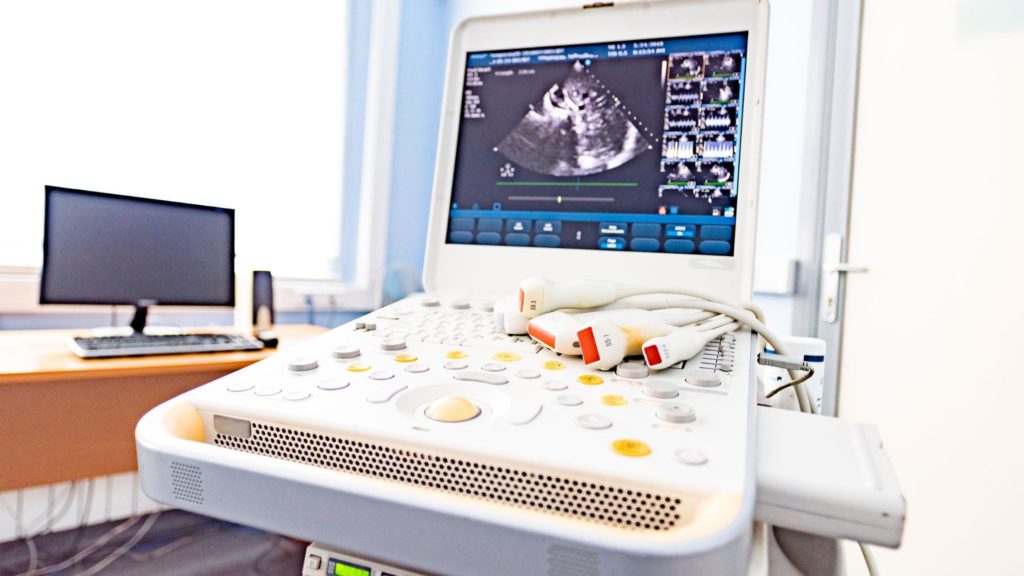
Key Components of an Ultrasound Machine

An ultrasound machine is a complex piece of medical equipment that relies on several crucial components to function effectively. Understanding these elements can help you appreciate the intricacies of this vital diagnostic tool.
Transducer Probe
The transducer probe is the heart of the ultrasound machine. It emits high-frequency sound waves and receives the echoes, converting them into electrical signals. This component comes in various shapes and sizes, each designed for specific examinations.
Central Processing Unit (CPU)
The CPU acts as the brain of the ultrasound machine, processing the electrical signals from the transducer and creating the images you see on the screen. It performs complex calculations to interpret the data and generate real-time visuals.
Display Monitor
The high-resolution monitor displays the processed ultrasound images. Modern machines often feature large, color LCD screens that provide clear, detailed views of the scanned area.
Control Panel
This interface allows technicians to adjust settings, input patient data, and manipulate images. It typically includes a keyboard, trackball, and various knobs and buttons for fine-tuning the ultrasound parameters.
Beam Former
The beam former focuses the ultrasound waves, improving image quality and resolution. It helps direct the sound waves to specific areas of interest within the body.
Image Processor
This component enhances the raw ultrasound data, applying filters and adjustments to produce clearer, more detailed images. It’s crucial for optimizing image quality and aiding in accurate diagnoses.
Data Storage System
Modern ultrasound machines include sophisticated storage systems that allow for saving, retrieving, and sharing patient images and data. This feature is essential for maintaining electronic health records and facilitating consultations.
Power Supply
A stable power supply ensures the consistent operation of the ultrasound machine. It regulates the electrical current to all components, maintaining the device’s performance and protecting it from power fluctuations.
Different Types of Ultrasound Machines
Ultrasound machines come in various types, each designed for specific medical applications. Here’s a detailed look at eight different types of ultrasound machines:
Portable Ultrasound Machines

These compact devices offer mobility and flexibility, making them ideal for point-of-care diagnostics. Portable ultrasound machines are battery-operated and can be easily transported between examination rooms or to patients’ bedsides. They’re particularly useful in emergency situations or rural healthcare settings.
Cart-Based Ultrasound Systems

These traditional, full-sized ultrasound machines offer superior image quality and advanced features. They’re typically found in hospitals and imaging centers, providing comprehensive diagnostic capabilities for a wide range of applications, from obstetrics to cardiology.
3D/4D Ultrasound Machines

These specialized devices produce three-dimensional images and real-time 3D videos (4D). They’re commonly used in obstetrics to provide detailed views of fetal development and in cardiology for assessing heart function.
Doppler Ultrasound Machines

Doppler ultrasound machines are designed to measure blood flow in vessels and organs. They’re crucial in diagnosing circulatory problems and assessing cardiovascular health.
Transvaginal Ultrasound Machines

These specialized devices use a probe inserted into the vagina to obtain detailed images of the female reproductive system. They’re particularly useful in early pregnancy assessments and diagnosing gynecological conditions.
Transrectal Ultrasound Machines

Used primarily for prostate examinations, these machines employ a probe inserted into the rectum to produce detailed images of the prostate gland and surrounding tissues.
Intravascular Ultrasound Machines

These highly specialized devices use a catheter-based system to provide detailed images of blood vessels from the inside. They’re invaluable in assessing arterial plaque buildup and guiding interventional procedures.
Veterinary Ultrasound Machines

Designed specifically for animal care, these ultrasound machines are adapted to the unique anatomical needs of various species. They’re used in veterinary clinics for diagnosing and monitoring a wide range of conditions in pets and livestock.
Common Medical Uses of Ultrasound Imaging
Ultrasound machines are versatile tools in modern medicine, offering non-invasive imaging for various diagnostic and therapeutic purposes. Here are seven common medical uses of ultrasound imaging:
Obstetrics and Fetal Monitoring
Ultrasound machines play a crucial role in monitoring fetal development during pregnancy. They allow healthcare providers to assess fetal growth, detect potential abnormalities, and determine the baby’s position and due date.
Cardiovascular Examinations
Cardiologists use ultrasound imaging to evaluate heart structure and function. This technique, known as echocardiography, helps diagnose heart valve problems, assess blood flow, and detect congenital heart defects.
Abdominal Imaging
Ultrasound is invaluable for examining abdominal organs such as the liver, gallbladder, pancreas, and kidneys. It aids in detecting gallstones, liver diseases, and kidney stones, among other conditions.
Musculoskeletal Evaluations
Orthopedic specialists utilize ultrasound to diagnose and assess soft tissue injuries, including tendon tears, muscle strains, and joint inflammation. This imaging modality is particularly useful for guiding therapeutic injections.
Breast Examinations
As a complement to mammography, ultrasound imaging helps differentiate between solid masses and fluid-filled cysts in breast tissue. It’s especially beneficial for examining dense breast tissue in younger women.
Thyroid Gland Assessment
Endocrinologists employ ultrasound to evaluate thyroid gland size, structure, and potential nodules. This non-invasive technique assists in diagnosing thyroid disorders and guiding fine-needle aspiration biopsies.
Vascular Studies
Ultrasound machines are essential for examining blood vessels, helping detect blood clots, assess arterial blockages, and evaluate blood flow in various parts of the body. This application is crucial in diagnosing conditions like deep vein thrombosis and peripheral artery disease.
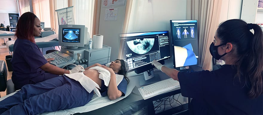
How is a Typical Ultrasound Performed?
Before your ultrasound machine examination, you may be asked to change into a hospital gown and remove any jewelry. Depending on the type of ultrasound, you might need to follow specific instructions, such as having a full bladder or fasting for several hours.
Positioning and Gel Application
You’ll be positioned on an examination table, either lying down or sitting up. The sonographer will apply a special water-based gel to the area being examined. This gel helps transmit the sound waves by eliminating air pockets between the transducer and your skin.
Scanning Process
The technician will then press the transducer firmly against your skin and move it back and forth over the area of interest. The ultrasound machine sends high-frequency sound waves into your body, which bounce off internal structures and return to the transducer.
Image Generation and Interpretation
As the sound waves are processed, real-time images appear on a nearby monitor. The sonographer may take still images or short video clips for further analysis. They might ask you to change positions or hold your breath briefly to obtain clearer images.
Completion and Clean-up
Once the examination is complete, the gel will be wiped off your skin. Most ultrasound procedures are painless and typically take between 30 minutes to an hour, depending on the complexity of the exam.
Results and Follow-up
After the ultrasound, the images are interpreted by a radiologist or your healthcare provider. You may receive preliminary results immediately, but a full report usually takes a few days. Your doctor will discuss the findings with you and recommend any necessary follow-up steps.
Benefits of Using Ultrasound for Medical Imaging and Diagnosing
The ultrasound machine has become an indispensable tool in modern medicine, offering numerous advantages for both patients and healthcare providers. Let’s explore seven key benefits of using ultrasound for medical imaging and diagnosing:
Non-Invasive and Painless
One of the most significant advantages of ultrasound imaging is its non-invasive nature. Unlike other imaging techniques that may require injections, incisions, or radiation exposure, ultrasound uses high-frequency sound waves to create images of internal structures. This means that patients can undergo ultrasound examinations without experiencing any pain or discomfort. The procedure is entirely external, making it an excellent option for those who may be anxious about more invasive diagnostic methods.
Furthermore, the non-invasive nature of ultrasound makes it particularly suitable for repeated examinations, allowing healthcare providers to monitor conditions over time without putting patients at risk.
Real-Time Imaging
Ultrasound machines provide real-time imaging capabilities, allowing medical professionals to observe organs and structures as they function. This dynamic visualization is invaluable for diagnosing a wide range of conditions and guiding certain medical procedures. For example, during a cardiac ultrasound, doctors can observe the heart’s chambers and valves in motion, providing crucial information about heart function and blood flow.
Real-time imaging also makes ultrasound an excellent tool for guiding minimally invasive procedures, such as biopsies or fluid drainage. The ability to see needle placement in real-time enhances accuracy and reduces the risk of complications.
No Radiation Exposure
Unlike X-rays or CT scans, ultrasound does not use ionizing radiation to produce images. This absence of radiation exposure makes ultrasound a safer option for patients, especially for those who require frequent imaging or are particularly sensitive to radiation, such as pregnant women and children.
The lack of radiation also means that there are no long-term risks associated with repeated ultrasound examinations, making it an ideal choice for monitoring chronic conditions or tracking fetal development throughout pregnancy.
Cost-Effective and Widely Available
Compared to other imaging modalities like MRI or CT scans, ultrasound machines are relatively inexpensive to purchase and operate. This cost-effectiveness translates to lower healthcare costs for patients and increased accessibility to diagnostic imaging services.
Moreover, the portability of many ultrasound machines allows for their use in various healthcare settings, from large hospitals to small clinics and even in remote or underserved areas. This widespread availability ensures that more patients can benefit from this valuable diagnostic tool.
Versatility in Applications
Ultrasound imaging is incredibly versatile, with applications across numerous medical specialties. From obstetrics and gynecology to cardiology, vascular medicine, and musculoskeletal imaging, ultrasound can be used to examine a wide range of body systems and structures.
This versatility extends to the types of information ultrasound can provide. Depending on the specific technique used, ultrasound can offer detailed information about organ structure, blood flow, tissue stiffness, and even cellular-level changes. This multifaceted capability makes ultrasound an invaluable tool for comprehensive patient assessment.
Immediate Results and Diagnosis
One of the most appreciated benefits of ultrasound is the immediacy of results. Unlike some imaging techniques that require extensive processing or interpretation time, ultrasound images are available instantly. This real-time feedback allows healthcare providers to make quick diagnosis and treatment decisions, potentially improving patient outcomes.
The immediacy of results also enhances the patient experience by reducing anxiety associated with waiting for test results. In many cases, doctors can discuss findings with patients immediately after the examination, allowing for prompt discussion of treatment options or further diagnostic steps if necessary.
Enhanced Patient Safety and Comfort
Ultrasound examinations are generally considered very safe, with no known harmful effects when used appropriately. This safety profile, combined with the non-invasive nature of the procedure, contributes to enhanced patient comfort and reduced anxiety during diagnostic imaging.
In conclusion, the ultrasound machine offers a unique combination of safety, versatility, and effectiveness that makes it an essential tool in modern healthcare. From its non-invasive nature and real-time imaging capabilities to its cost-effectiveness and wide-ranging applications
Disadvantages of Ultrasound Procedures
While ultrasound machines have revolutionized medical imaging and diagnostics, it’s important to consider their limitations and potential drawbacks. Understanding these disadvantages can help healthcare providers and patients make informed decisions about when to use ultrasound procedures and when alternative imaging methods might be more appropriate.
Limited Penetration and Image Quality
One of the primary disadvantages of ultrasound procedures is their limited ability to penetrate deep into the body. The ultrasound machine relies on sound waves to create images, and these waves can only travel so far before losing strength and clarity. This limitation becomes particularly evident when attempting to image structures that are far from the body’s surface or hidden behind bone or gas-filled organs.
For instance, ultrasound may struggle to provide clear images of:
- Deep abdominal organs in individuals with a high body mass index (BMI)
- Structures within or behind the lungs, as air interferes with sound wave transmission
- Brain tissue, due to the skull’s density
Additionally, the quality of ultrasound images can be affected by various factors, including:
- Patient movement during the procedure
- The skill and experience of the ultrasound technician
- The specific type and capabilities of the ultrasound machine being used
These limitations in image quality and penetration may necessitate the use of other imaging modalities, such as CT scans or MRIs, in certain situations.
Operator Dependence
Another significant disadvantage of ultrasound procedures is their heavy reliance on the skill and experience of the operator. Unlike some other imaging techniques that produce standardized images, ultrasound requires real-time manipulation of the transducer to obtain the best possible views of the target structures.
This operator dependence can lead to several issues:
- Variability in image quality and interpretation between different technicians
- Potential for missed diagnoses if the operator fails to capture all relevant views
- Challenges in reproducing exact images for follow-up comparisons
To mitigate these risks, healthcare facilities must invest in thorough training programs and ongoing education for their ultrasound technicians. However, even with extensive training, some degree of variability will always exist due to the nature of the technology.
Limited Field of View
Ultrasound machines typically provide a relatively narrow field of view compared to other imaging modalities. This limitation can make it challenging to:
- Visualize large anatomical structures in their entirety
- Detect diffuse pathologies that affect a broad area
- Provide comprehensive imaging of complex anatomical regions
For example, while an ultrasound may be excellent for examining a specific area of the liver, it may struggle to provide a complete overview of the entire organ and its surrounding structures. This narrow field of view can sometimes lead to incomplete diagnoses or the need for multiple scans to fully assess an area of concern.
Difficulty Imaging Certain Tissues and Structures
Some types of tissue and anatomical structures pose particular challenges for ultrasound imaging. For instance:
- Bone: Ultrasound waves cannot penetrate bone effectively, limiting its use in orthopedic imaging beyond surface-level structures.
- Gas-filled organs: The presence of gas, such as in the lungs or intestines, can create artifacts and distortions in ultrasound images.
- Calcifications: Dense calcium deposits can block ultrasound waves, creating shadows in the image that may obscure underlying structures.
These limitations can be particularly problematic when trying to diagnose conditions that affect these types of tissues or when attempting to visualize structures located near or behind them.
Potential for Misdiagnosis
The combination of operator dependence, limited penetration, and image quality issues can sometimes lead to misdiagnosis or missed diagnoses. This risk is particularly concerning in critical care situations or when screening for serious conditions such as cancer.
Factors that can contribute to misdiagnosis include:
- Misinterpretation of normal anatomical variations as pathological findings
- Failure to detect small or subtle abnormalities due to image quality limitations
- Overreliance on ultrasound when other imaging modalities might be more appropriate
To mitigate this risk, healthcare providers often use ultrasound in conjunction with other diagnostic tools and clinical assessments, rather than relying on it as a standalone diagnostic method.
Time-Consuming Nature of Some Procedures
While many ultrasound examinations are relatively quick, certain specialized procedures can be time-consuming. For example:
- Comprehensive echocardiograms may take up to an hour to complete
- Detailed musculoskeletal examinations can require extensive
Frequently Asked Questions
Here are some frequently asked questions and answers about an Ultrasound machine.
Are ultrasound machines safe?
Yes, ultrasound machines are generally considered very safe. Unlike X-rays or CT scans, ultrasound does not use ionizing radiation, which can be harmful in large doses. Instead, it uses sound waves, which have not been shown to cause any harmful effects when used for medical imaging.
However, as with any medical procedure, ultrasounds should only be performed when medically necessary and by trained professionals. The FDA recommends against non-medical use of ultrasound, such as for “keepsake” images or videos during pregnancy.
Can ultrasound replace other imaging methods like CT or MRI?
No, ultrasound cannot completely replace other imaging modalities like CT or MRI. Each imaging method has its strengths and weaknesses, and they often complement each other in diagnostic workflows. Ultrasound excels at real-time imaging of soft tissues, blood flow, and certain anatomical structures, but it has limitations in penetration, field of view, and imaging certain tissues like bone or air-filled organs. CT and MRI provide superior detail and cross-sectional imaging capabilities, making them more suitable for certain applications.
Are there any contraindications for ultrasound imaging?
Ultrasound is generally considered safe for most patients, but there are a few contraindications or situations where caution is warranted:
- During the first trimester of pregnancy, some healthcare providers may limit or avoid certain types of ultrasound examinations as a precautionary measure.
- Patients with certain implanted devices, such as pacemakers or defibrillators, may require special precautions or alternative imaging methods due to potential interference from ultrasound waves.
- In some cases, ultrasound may be contraindicated for patients with recent surgical incisions or open wounds in the area being examined, as the pressure from the transducer could cause discomfort or disrupt healing.
Healthcare providers will evaluate each patient’s specific circumstances and weigh the potential risks and benefits before proceeding with an ultrasound examination.
How does an ultrasound machine work?
An ultrasound machine works by emitting high-frequency sound waves into the body and then capturing the echoes as they bounce back. Here’s a simplified explanation of the process:
- The transducer (probe) sends out sound waves
- These waves travel through body tissues
- Different tissues reflect the waves differently
- The transducer picks up the reflected waves
- The machine’s computer processes these echoes
- An image is generated on the monitor
The ultrasound machine’s sophisticated software interprets the time it takes for echoes to return and the strength of these echoes to create detailed images of structures within the body.
How often do ultrasound machines need maintenance?
Regular maintenance is crucial for ensuring the accuracy and longevity of ultrasound machines. Typically, manufacturers recommend:
- Daily: Basic cleaning and visual inspection
- Weekly: More thorough cleaning and system checks
- Annually: Comprehensive service by a qualified technician
The exact maintenance schedule may vary depending on the specific model and frequency of use. It’s essential to follow the manufacturer’s guidelines to keep the ultrasound machine in optimal condition.
Can ultrasound machines be used for therapeutic purposes?
Yes, ultrasound machines are not only used for diagnostic imaging but also for therapeutic applications. Some therapeutic uses include:
- Physical therapy: To promote healing in soft tissues and reduce pain
- Breaking up kidney stones: High-intensity focused ultrasound (HIFU) can be used to fragment kidney stones
- Cancer treatment: HIFU is being explored as
Conclusion
As you’ve learned, ultrasound machines are sophisticated devices that use sound waves to create images of internal body structures. By understanding how these machines work, you can better appreciate their critical role in medical diagnostics and patient care.
Whether you’re a healthcare professional or simply curious about medical technology, recognizing the principles behind ultrasound imaging enhances your knowledge of this vital tool. As ultrasound technology continues to advance, it will undoubtedly play an even greater role in medicine, offering improved image quality and expanded applications. By staying informed about developments in this field, you’ll be well-equipped to navigate the evolving landscape of medical imaging and its impact on healthcare.
Discussion about this post